Brian Howe DDS, Family Dentistry | Emergency Treatment, Laser Dentistry and All-on-6 reg

CBCT

At the office of Brian Howe DDS, Family Dentistry, we rely on advanced imaging to guide precise, predictable care. Cone-beam computed tomography (CBCT) gives our team three-dimensional views of the teeth, jaws, and surrounding anatomy that conventional two-dimensional X-rays cannot capture. Those clear, volumetric images help clinicians identify subtle problems, plan complex procedures, and tailor treatment to each patient’s unique anatomy.

Our approach combines the clinical judgment of experienced dentists with the objective detail provided by CBCT scans. The goal is straightforward: deliver safer, more efficient care with fewer surprises and better long-term outcomes. Below you’ll find an overview of what CBCT is, why it matters, how we use it across specialties, and what patients can expect when they come in for a scan.

How CBCT differs from traditional dental X-rays

Traditional dental X-rays produce flat images that are valuable for many routine screenings, but they can obscure overlapping structures and miss depth-related information. CBCT acquires a cone-shaped beam while rotating around the head, creating a volumetric dataset that can be viewed from any angle. That third dimension reveals relationships between teeth, bone, nerves, and sinuses with a clarity impossible on two-dimensional films.

This imaging modality is particularly useful when clinicians need to measure distances, evaluate bone volume, or inspect the internal structure of roots and surrounding tissues. Rather than interpreting shadows, dentists can examine true spatial relationships, which supports more predictable surgical and restorative results. For patients, that means treatment plans are based on direct visualization rather than inference.

Although CBCT provides much more detail, it is used selectively where the extra information will meaningfully influence care. Our team considers the diagnostic question first and chooses CBCT when the benefits of three-dimensional imaging justify its use. That careful, case-by-case approach ensures patients receive thoughtful, evidence-based imaging tailored to their needs.

Planning dental implants and complex surgery with exacting detail

One of CBCT’s most transformative uses is in implant dentistry. Successful implant placement depends on knowing exactly how much bone is available, where vital structures like nerves and sinuses are located, and the ideal angulation for each implant. CBCT scans provide precise measurements and cross-sectional views that let clinicians plan implant size, position, and orientation before a single incision is made.

With this information, our team can reduce surgical guesswork, minimize the risk of complication, and often shorten chair time during the actual procedure. In many cases, CBCT data is used to fabricate surgical guides that translate a virtual plan into an accurate clinical outcome. The result is a more predictable process from planning through healing.

CBCT is also invaluable for other surgical procedures—such as impacted tooth removal, bone grafting, and complex extractions—where understanding root relationships and adjacent anatomy is critical. Enhanced visualization improves safety and helps the care team choose the least invasive approach that still achieves the desired clinical goals.

Sharper diagnostics for endodontics, orthodontics, and airway assessment

Beyond implant planning, CBCT supports several specialty diagnoses. In endodontics, three-dimensional imaging can reveal accessory canals, root fractures, or persistent infections that are not visible on conventional films. That level of detail supports more targeted retreatment or surgical decisions when conventional imaging falls short.

In orthodontics and restorative planning, CBCT helps assess jaw relationships, impacted teeth, and skeletal asymmetries. Orthodontists and dentists can visualize tooth position in three planes and evaluate how proposed movements will interact with surrounding tissues. That foresight improves outcomes and can reduce treatment time by avoiding unexpected obstacles.

CBCT is also used for airway assessment when sleep-disordered breathing or airway obstruction is a concern. The scans allow clinicians to evaluate airway volume and anatomical contributors to obstruction, providing an evidence-based starting point for discussions about therapy and referrals when multidisciplinary care is appropriate.

Safety, radiation considerations, and patient comfort

Patients often ask about radiation exposure with advanced imaging. Modern CBCT units are designed to limit dose through collimation and adjustable fields of view so only the necessary anatomy is scanned. When compared to medical CT scans, dental CBCT typically exposes patients to substantially lower radiation levels, especially when small-volume scans are used for localized questions.

Our office follows the principle of ALARA—keeping radiation As Low As Reasonably Achievable—by selecting the smallest effective field of view and the appropriate resolution for each case. We also document the diagnostic need for each scan and discuss it with patients, ensuring that imaging is justified and purposeful.

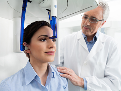
The scanning process itself is quick and comfortable: most CBCT scans take less than a minute to acquire. Patients remain seated or standing with minimal movement, and there is no need for intravenous contrast or complex preparation. This streamlined workflow makes CBCT a practical option when three-dimensional information will affect diagnosis or treatment planning.

From images to action: how scans become better treatment

A CBCT scan is only as useful as the interpretation and the way it’s integrated into care. Our clinicians review the datasets in three dimensions, taking measurements, assessing bone quality, and noting anatomical variations that may influence treatment. The images are then correlated with clinical findings, intraoral photographs, and conventional radiographs to form a comprehensive, patient-specific plan.

In many cases, the CBCT data is incorporated into digital workflows—merging with intraoral scans, case-planning software, or surgical guide fabrication—to translate virtual plans into precise clinical steps. This integration helps reduce surprises in the operatory and supports smoother restorative and surgical phases.

Equally important is communication: we take time to explain what the images show and how they support proposed treatment options. Patients receive clear, jargon-free explanations so they can make informed decisions about their care and understand the rationale behind recommended procedures.

Expertise, quality control, and ongoing care

Proper use of CBCT requires training in both image acquisition and interpretation. Our clinicians have experience reading three-dimensional datasets and applying findings directly to clinical cases. That experience matters because misinterpretation can lead to unnecessary procedures or missed diagnoses. We maintain a rigorous approach to image review and consult with specialists when complex or unusual findings arise.

Quality control is part of every scan: positioning, exposure settings, and artifact reduction are monitored to ensure diagnostic-grade images. When additional input is needed—such as from oral and maxillofacial radiology or other dental specialists—we coordinate care to ensure patients benefit from a multidisciplinary perspective.

CBCT is a diagnostic tool that supports ongoing, informed care. Whether a scan is used to guide immediate treatment or to monitor healing and long-term outcomes, the three-dimensional record it creates becomes part of a patient’s dental history and helps guide future decisions.

In summary, cone-beam computed tomography provides a meaningful upgrade in diagnostic capability when three-dimensional information will change the course of care. By combining experienced clinicians, careful case selection, and modern imaging technology, Brian Howe DDS, Family Dentistry uses CBCT to improve accuracy, safety, and predictability across a range of dental treatments. If you have questions about whether CBCT is appropriate for your situation or would like to learn more about our imaging protocols, please contact us for more information.

Frequently Asked Questions

What is cone-beam computed tomography (CBCT)?

+

Cone-beam computed tomography, commonly called CBCT, is a three-dimensional dental imaging technique that captures volumetric images of the teeth, jaws and surrounding anatomy. At Brian Howe DDS, Family Dentistry, we use CBCT to visualize spatial relationships that two-dimensional X-rays cannot show, such as the exact position of roots, bone contours and adjacent sinuses. The scan acquires a cone-shaped beam while rotating around the head, creating a dataset that can be reformatted into cross-sections, panoramic views and 3D renderings for clinical review.

CBCT is a diagnostic tool rather than a treatment; its primary value lies in revealing detailed anatomy to guide decisions. Clinicians can measure distances, assess bone volume and inspect internal tooth structure directly rather than inferring from shadows. When selected appropriately, CBCT provides actionable information that improves diagnostic confidence and supports more predictable care.

How does CBCT differ from traditional dental X-rays?

+

Traditional dental X-rays produce flat images that are excellent for many routine screenings but can obscure overlapping structures and depth information. CBCT generates a volumetric dataset that can be viewed from any angle, revealing true spatial relationships between teeth, bone, nerves and sinuses. That additional dimension is especially helpful when clinicians must measure bone thickness, evaluate root anatomy or locate critical structures prior to surgery.

Because of its enhanced detail, CBCT is used selectively rather than as a replacement for conventional films. Two-dimensional X-rays remain the first-line option for many routine checks and bitewing exams, while CBCT is reserved for cases where three-dimensional information will meaningfully influence diagnosis or treatment planning. This approach balances diagnostic benefit with prudent use of imaging resources.

When is CBCT recommended for dental patients?

+

CBCT is recommended when the clinical question requires three-dimensional visualization to reduce uncertainty or risk. Typical indications include implant planning, evaluation of impacted or ectopic teeth, complex extractions, assessment of suspected root fractures or persistent endodontic infections, and airway analysis for suspected sleep-disordered breathing. The decision to scan is made case by case, with clinicians weighing how the information will affect treatment choices and outcomes.

Our team follows evidence-based guidelines to determine when CBCT will add value to a case. We document the diagnostic need, select the smallest effective field of view and discuss the rationale with patients so they understand how the scan supports recommended care. This selective strategy ensures imaging is purposeful and clinically justified.

How does CBCT improve dental implant planning and surgical accuracy?

+

CBCT provides precise measurements of bone height, width and density and shows the position of vital structures such as the inferior alveolar nerve and maxillary sinuses. With these cross-sectional views clinicians can select proper implant size, angulation and position before surgery, reducing guesswork and lowering the risk of complications. The dataset can also be used to design and fabricate surgical guides that translate a virtual plan into consistent intraoperative placement.

Using CBCT for implant cases often shortens operative time and improves predictability during the restorative phase. By identifying anatomic constraints or the need for grafting in advance, the surgical team can plan steps more efficiently and communicate expected outcomes to the patient. This preoperative detail helps create a smoother transition from planning to restoration and healing.

Can CBCT detect root fractures, accessory canals or persistent infections?

+

Yes. CBCT excels at revealing features that are difficult or impossible to see on two-dimensional radiographs, including vertical root fractures, accessory canals and localized areas of bone loss from infection. In endodontics, three-dimensional imaging can change the diagnosis or treatment plan by revealing the true extent and location of pathology. That level of detail supports more targeted retreatment, surgical approaches or referrals when necessary.

Interpreting these findings requires experience, so clinicians correlate CBCT images with clinical tests and conventional radiographs before finalizing a plan. When findings are complex or ambiguous, consultation with an endodontist or oral and maxillofacial radiologist helps ensure appropriate interpretation. The result is more accurate diagnoses and better-informed decisions about therapy.

How is CBCT used in orthodontic and airway assessments?

+

In orthodontics, CBCT allows clinicians to evaluate tooth position in three planes, assess impacted teeth and identify skeletal asymmetries that influence treatment strategy. The scans help visualize root proximity, cortical bone boundaries and the relationship between jaws, which can inform decisions about extractions, surgical interventions or mechanics. This spatial information improves planning for complex movements and helps anticipate potential obstacles during active treatment.

For airway assessment, CBCT can quantify airway volume and highlight anatomic contributors to obstruction, such as constricted nasal passages or retroglossal narrowing. These objective measures provide a starting point for discussions about sleep-disordered breathing and possible referrals to sleep medicine or ENT specialists. CBCT is one component of a comprehensive evaluation and is interpreted alongside clinical examination and sleep history.

What should patients expect during a CBCT appointment?

+

A CBCT appointment is generally quick and straightforward: patients remain seated or standing while the scanner rotates around the head and acquires images in under a minute. There is no need for intravenous contrast or special preparation, and most patients experience minimal discomfort during positioning. Staff will explain the process, ensure proper stabilization to reduce motion and select the appropriate field of view for the clinical question.

Patients should inform the team of pregnancy or recent medical imaging so the staff can follow appropriate precautions. After acquisition, the images are reviewed by the clinician and incorporated into the diagnostic workflow; staff will discuss the findings and next steps in clear, nontechnical language. The overall appointment is designed to be efficient while producing high-quality diagnostic information.

Is CBCT safe and what are the radiation considerations?

+

CBCT exposes patients to more radiation than a single dental bitewing but substantially less than most medical CT studies when appropriate protocols are used. Our office follows the principle of ALARA—As Low As Reasonably Achievable—by selecting the smallest effective field of view and the appropriate resolution for each diagnostic task. These measures minimize dose while preserving the image quality needed for accurate interpretation.

Clinicians always document the diagnostic justification for a scan and will discuss risks and benefits with patients when imaging is proposed. When alternative imaging can answer the clinical question, conventional radiographs remain the preferred option. This careful, evidence-based approach ensures that CBCT is used only when the additional three-dimensional information will meaningfully affect care.

How are CBCT images interpreted and integrated into treatment plans?

+

Interpreting CBCT datasets involves reviewing multiplanar views, taking precise measurements and correlating findings with the clinical exam and conventional radiographs. Clinicians use software tools to assess bone quality, measure distances to critical structures and simulate implant positions or surgical approaches. When appropriate, CBCT images are merged with intraoral scans and planning software to create surgical guides and digital treatment workflows.

Clear communication is an important part of integration: clinicians explain what the images reveal and how they influence recommended options so patients can make informed decisions. For complex cases the practice coordinates input from specialists or oral and maxillofacial radiologists to ensure comprehensive interpretation. This multidisciplinary approach helps translate imaging into safe, predictable clinical steps.

Who performs and reviews CBCT scans at the practice?

+

CBCT acquisition is performed by trained clinical staff under the supervision of licensed dentists, and image review is carried out by clinicians with experience interpreting three-dimensional datasets. At the office of Brian Howe DDS, Family Dentistry, team members follow quality control protocols for positioning, exposure settings and artifact reduction to ensure diagnostic-grade images. When scans reveal complex or unfamiliar findings, we consult with specialists or oral and maxillofacial radiologists to provide additional expertise.

Maintaining proficiency in both image acquisition and interpretation is essential for safe and effective use of CBCT. The practice documents each scan, keeps it in the patient record and uses the dataset as part of ongoing care and monitoring. This structured workflow ensures that CBCT contributes meaningfully to treatment planning and long-term outcomes.

Contact Us

Our friendly and knowledgeable team is always ready to assist you. You can reach us by phone at (740) 393-2161 or by using the convenient contact form below. If you submit the form, a member of our staff will respond within 24–48 hours.

Please do not use this form for emergencies or for appointment-related matters.

SMS Consent
Submit

Thank You!

We appreciate you taking the time to visit our site. We'll review your message and be in touch with you soon.

Continue
Brian Howe DDS, Family Dentistry | Oral Exams, All-on-4 reg  and Digital Impressions