Existing Patients
(740) 393-2161
New Patients
(740) 200-4777

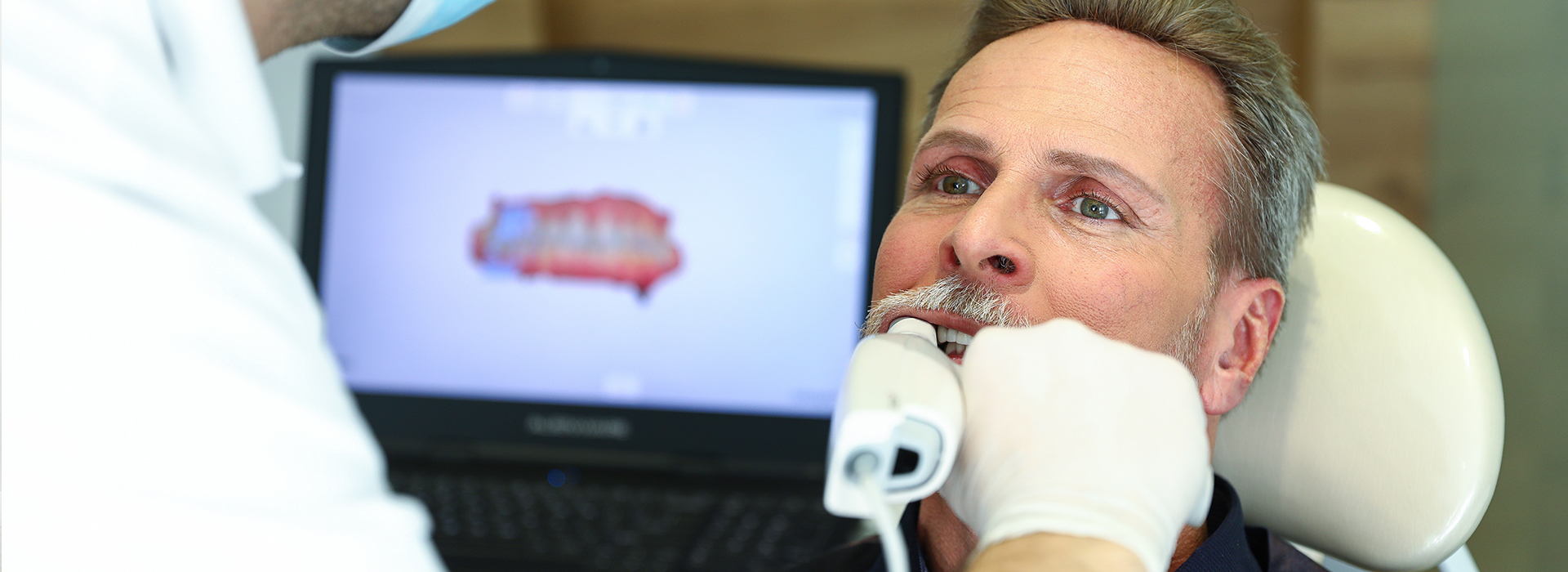
Digital impressions use small, handheld intraoral scanners to capture a precise, three-dimensional image of your teeth and surrounding oral tissues. Unlike traditional putty impressions, the scanner records thousands of data points in real time, creating an accurate computer-generated model that dentists and dental technicians can use immediately. This process preserves fine details—occlusal surfaces, margins, and soft-tissue contours—so restorations and appliances fit more predictably.
For patients, the practical benefit is straightforward: the reading is fast, predictable, and far less invasive than conventional methods. For clinicians, the data can be manipulated on-screen to evaluate occlusion, check margins, and communicate specific adjustments to the dental laboratory. The result is a workflow built on clarity and repeatability rather than guesswork and manual remakes.
At Brian Howe DDS, Family Dentistry, we integrate digital impressions into routine restorative and prosthetic care because they reduce variability and improve clinical outcomes. Whether preparing a crown, designing an implant restoration, or capturing records for orthodontic planning, digital models provide a dependable foundation for treatment decisions.
One of the most noticeable differences patients report is the level of comfort. Traditional impressions often require bulky trays and impression material that can trigger gagging or prolonged mouth-holding. Digital scanning eliminates those elements: the scanner tip is compact and moves quickly, limiting discomfort and enabling scans for patients of almost any age or sensitivity level.
Precision comes from both the technology and the operator. Modern scanners produce high-resolution images that reveal microscopic detail, and when used by a trained clinician, they capture full-arch data with consistent accuracy. This precision matters most when laboratory technicians use the digital file to fabricate restorations—accurate scans reduce the need for adjustments and result in better-fitting crowns, bridges, and appliances.
Another practical advantage for patients is documentation. Digital models become part of the patient’s chart and can be referenced at any time—useful for monitoring wear, planning future work, or comparing pre- and post-treatment anatomy without repeating uncomfortable procedures.
Once a scan is complete, the digital file can be transmitted electronically to a dental lab or sent to an in-office milling system. This digital transfer eliminates postal delays, minimizes handling errors, and preserves the integrity of the impression data. Labs receive a standardized, high-fidelity dataset that speeds production and helps technicians deliver restorations with fewer adjustments.
The digital workflow also introduces quality checks earlier in the process. Technicians and clinicians can review the scan together, identify potential issues, and request additional data before fabrication begins. That collaboration reduces the likelihood of remakes and shortens the overall treatment timeline, which benefits both the patient and the practice.
From a clinical standpoint, electronic records simplify case management. Files can be archived, duplicated, and integrated with other digital tools—such as implant planning software or CAD/CAM systems—so restorative planning is more coordinated and traceable from the initial scan to final delivery.
Digital impressions are a critical component of same-day, chairside dentistry. By pairing the scan with CAD/CAM milling, clinicians can design, mill, and place ceramic restorations within a single appointment. This model reduces the number of visits required for patients and eliminates temporary restorations in many cases, improving both convenience and comfort.
Chairside systems rely on accurate digital impressions to produce restorations that meet aesthetic and functional demands. Because the data capture is immediate, clinicians can make on-the-spot adjustments to the digital design, preview how a restoration will sit in occlusion, and refine margins before milling begins. The in-office cycle—from scan to final polish—delivers a controlled, efficient experience for patients who prefer to complete treatment quickly.
Beyond same-day crowns, the chairside workflow supports a range of applications including inlays, onlays, and certain prosthetic components. For complex cases, a hybrid approach—combining in-office scanning with laboratory fabrication—offers flexibility while keeping the advantages of digital precision.
Preparing for a digital impression is usually simple: routine hygiene and the removal of excessive debris make scans faster and cleaner. The clinician may recontour soft tissue or use a gentle retraction technique to expose margins clearly when necessary. Because the scanner captures both hard and soft tissues, careful preparation helps ensure the final model reflects the true treatment area.
After the scan, the digital file is reviewed to confirm completeness and clarity. Small rescans can be performed immediately if an area needs better detail, avoiding the delays associated with traditional retakes. Once approved, the file is either sent to a trusted laboratory partner or used within the office for CAD/CAM fabrication.
Post-delivery care follows standard restorative protocols: clinicians will check fit, occlusion, and aesthetics, make any minor adjustments, and provide guidance on home care. Because a digital record exists, follow-up visits are informed by precise baseline data, which helps the team monitor restoration performance and address concerns efficiently.
Digital impressions represent a meaningful advancement in restorative and prosthetic dentistry, improving comfort for patients and predictability for clinicians. If you’d like to learn more about how our digital workflows support crowns, bridges, implant restorations, and same-day options, please contact us to discuss your treatment goals with a member of our team.
Digital impressions use small intraoral scanners to capture a detailed three-dimensional record of teeth and surrounding soft tissues in real time. The scanner records thousands of data points to produce a computer-generated model that preserves occlusal surfaces, margins and soft-tissue contours for predictable restorative work. This digital dataset replaces bulky impression trays and creates a more repeatable foundation for laboratory and chairside procedures.
Clinicians can manipulate digital models on-screen to evaluate occlusion, check margins and plan restorations with greater clarity and consistency. At Brian Howe DDS, Family Dentistry we integrate digital impressions into restorative and prosthetic workflows to reduce variability and improve clinical outcomes. Whether used for crowns, implant restorations or orthodontic records, digital models support more informed treatment decisions.
Traditional impressions rely on impression trays and elastomeric materials that set around the dentition, while digital impressions are captured electronically with a handheld scanner. The digital approach eliminates the need for impression material, reduces gag reflex triggers and often shortens procedure time. Because the scan is produced instantly, clinicians can review the capture immediately and perform targeted rescans when needed.
From a technical standpoint, digital files are standardized and transferable, which reduces handling errors associated with physical models. Labs receive high-fidelity datasets that help technicians fabricate restorations with fewer adjustments. The digital workflow also allows for easier integration with CAD/CAM design and implant planning software for more coordinated case management.
Modern intraoral scanners produce high-resolution images capable of capturing microscopic details relevant to crown margins, contact areas and occlusal anatomy. Accuracy depends on the scanner technology and the operator’s technique, but for single-unit and short-span restorations digital scans commonly achieve clinically acceptable precision. Careful scanning protocols and operator training are important to ensure consistent, reproducible results.
For implant restorations, digital impressions capture implant scan bodies and surrounding tissues to create an accurate virtual model for lab fabrication or in-office CAD/CAM workflows. When clinicians and technicians review the digital file together, early detection of potential concerns reduces adjustments and helps deliver restorations that fit more predictably. In complex full-arch cases, clinicians may combine digital data with other records to ensure comprehensive accuracy.
Patients typically report that digital scanning is more comfortable than traditional impressions because it eliminates bulky trays and setting material. The scanner tip is compact and moves quickly, which minimizes prolonged mouth opening and reduces the likelihood of gagging or discomfort. Scans are generally completed in a matter of minutes depending on the scope of the case and the area being captured.
Another practical benefit is that rescans can be performed immediately if an area needs better detail, avoiding the delays and discomfort of retaking physical impressions. The resulting digital models become part of the patient chart and can be referenced for future care, monitoring wear or comparing pre- and post-treatment anatomy. Overall, the process tends to be faster, less invasive and easier to document.
Yes. Once a scan is complete the digital file can be transmitted electronically to a dental laboratory or sent to an in-office milling system, which eliminates postal delays and minimizes handling errors. Labs receive a standardized dataset with high fidelity that speeds production and supports more accurate fabrication. Electronic transfer preserves the integrity of the impression data and reduces the chance of distortion inherent to physical impressions.
The digital workflow also enables collaborative quality checks early in the process so technicians and clinicians can identify issues and request additional data before fabrication begins. This collaboration reduces the likelihood of remakes and shortens the overall treatment timeline. For practices, fewer remakes mean improved efficiency and a more reliable patient experience.
Digital impressions are a key component of same-day dentistry because they provide an immediate, accurate digital model that can be used with CAD/CAM design and milling systems. After scanning, clinicians can design a restoration on-screen, make adjustments to occlusion and margins, and send the design to an in-office mill for fabrication. This sequence allows clinicians to deliver ceramic restorations within a single appointment in many cases, eliminating the need for temporaries and additional visits.
Chairside systems also support a hybrid approach where complex cases are planned digitally but finished in a laboratory, providing flexibility when needed. At Brian Howe DDS, Family Dentistry we use chairside and lab-based digital workflows to match clinical needs and patient preferences. Immediate visualization and on-the-spot adjustments give clinicians greater control over fit and aesthetics before milling begins.
Preparing for a digital impression is straightforward: routine hygiene and the removal of excessive debris make scans faster and clearer. Clinicians may use gentle retraction or recontouring of soft tissue to expose margins when necessary, and dry fields help the scanner capture accurate detail. Clear communication from the dental team about what to expect reduces anxiety and speeds the appointment.
After the scan the digital file is reviewed for completeness and clarity, and small rescans can be performed on the spot if needed. Once approved, the file is either sent to a trusted laboratory partner or used in-office for CAD/CAM fabrication, and the digital model is archived in the patient’s chart. Post-delivery care follows standard restorative protocols, with follow-up visits informed by precise baseline data captured during the scan.
Digital impressions are noninvasive and use optical scanning rather than radiation, making them safe for patients of all ages when performed properly. The compact scanner tip and brief capture times make scans well suited to children, patients with strong gag reflexes, and those with limited tolerance for lengthy procedures. Clinicians adjust their technique and pacing to accommodate sensitivity and to ensure patient comfort throughout the appointment.
There are occasional situations—such as severe bleeding, extensive soft-tissue inflammation or very limited mouth opening—where a clinician may recommend an alternate approach. In those cases the dental team will evaluate the clinical circumstances and choose the best method to produce a reliable impression while prioritizing the patient’s comfort and the integrity of the restoration.
Digital impressions offer many advantages but are not universally ideal for every situation. Challenges can arise with subgingival margins that are difficult to access, extremely reflective or translucent materials that interfere with optical scanning, and complex full-arch edentulous cases where some clinicians still prefer conventional techniques. Operator experience and case complexity influence whether a digital scan alone will meet clinical needs.
In practice, many clinicians use a hybrid approach: they capture what can be recorded digitally and combine that information with traditional methods or laboratory techniques when warranted. The decision to use digital or conventional impressions is made on a case-by-case basis to ensure the most accurate, durable and functional outcome for the patient.
Digital scan files are typically archived in the practice’s secure record system and can be duplicated, backed up and integrated with other digital tools such as implant planning and CAD/CAM software. Having an archived digital model allows clinicians to compare anatomy over time, monitor wear and document baseline conditions without repeating uncomfortable procedures. The portability of digital files also facilitates communication with laboratories and specialty colleagues during treatment planning.
Access controls, backups and standardized file formats help protect the integrity and availability of scan data for future use. Clinicians can combine archived scans with radiographic records and surgical guides to create coordinated, traceable plans from initial scan to final restoration. Secure storage and interoperability therefore enhance long-term case management and clinical collaboration.
Our friendly and knowledgeable team is always ready to assist you. You can reach us by phone at (740) 393-2161 or by using the convenient contact form below. If you submit the form, a member of our staff will respond within 24–48 hours.
Please do not use this form for emergencies or for appointment-related matters.
