Brian Howe DDS, Family Dentistry | Cosmetic Dentistry, Sleep Apnea and Digital Impressions

Digital Radiography

Digital radiography: a modern foundation for dental imaging

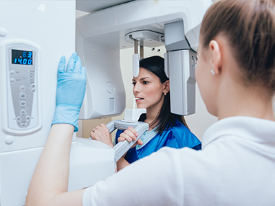
Digital radiography has become the standard for diagnostic imaging in contemporary dental care. At Brian Howe DDS, Family Dentistry, we use digital X‑ray systems that capture high-resolution images quickly and reliably, supporting accurate treatment planning and clear communication with patients. Unlike older film techniques, digital imaging streamlines the entire process—from acquisition to storage—so clinicians can focus more time on diagnosis and patient care.

These systems are designed to integrate with electronic patient records, allowing images to be attached directly to your chart the moment they’re taken. That integration helps our team track changes over time, compare past and present images, and make informed recommendations during appointments. For patients, the result is a more transparent visit: you can see what we see and better understand the rationale behind suggested treatments.

Beyond convenience, digital radiography supports a higher standard of care by making images easier to enhance, share, and archive. This technology helps our clinicians spot subtle issues earlier and coordinate with specialists when necessary, which contributes to more predictable outcomes and a smoother patient experience.

Inside the system: sensors, software, and seamless records

At the heart of digital radiography are compact electronic sensors that replace traditional film. These sensors capture X‑ray information and convert it into a digital file almost instantly. Once captured, the image is routed into secure practice software where it becomes part of the patient’s record—accessible to the dentist during the same visit and preserved for future comparison.

Modern imaging software gives clinicians tools to enhance contrast, zoom into trouble spots, and measure structures with precision. Those tools make it easier to evaluate decay, check the fit of restorations, or monitor bone levels without waiting for film to be developed. Because images are digital, they can be viewed simultaneously by more than one clinician, shared with a specialist for consultation, or included in diagnostic workflows such as implant planning or endodontic assessment.

From a record‑keeping perspective, digital files simplify organization and retrieval. They reduce the risk of lost or damaged films and make it straightforward to compile complete diagnostic histories. Secure backups and standardized formats help ensure images remain available and readable over time, supporting continuity of care across years and different providers.

Radiation safety: meaningful reductions and careful protocols

One of the most important advantages of digital radiography is its potential to lower patient exposure to ionizing radiation. Because digital sensors are more sensitive than conventional film, clinicians can obtain diagnostically useful images using less radiation in many cases. This contributes to safer imaging practices while preserving the detail necessary for accurate diagnosis.

Safety is always a priority in our imaging workflow. Along with using efficient digital sensors, we follow established protective measures such as appropriate collimation, correct exposure settings, and the selective use of X‑rays based on each patient’s clinical needs. Protective shielding and standard positioning techniques are used as appropriate to minimize unnecessary exposure for every patient.

For patients who are pregnant, very young, or have particular concerns, our team discusses imaging options and tailors the approach to balance diagnostic value and safety. The goal is to obtain the information needed for high‑quality care while using the least amount of radiation reasonably possible.

Sharper images, faster diagnoses, and clearer patient communication

Digital images often reveal subtle details that help clinicians detect early decay, assess periodontal bone levels, and evaluate the status of existing restorations. The ability to enhance contrast, adjust brightness, and magnify areas of interest can make it easier to identify problems that might be missed on lower‑quality film. This leads to more confident diagnoses and better-informed treatment recommendations.

Because images are available immediately, the diagnostic discussion can take place during the same appointment. Clinicians can show patients the image on a screen, point out areas of concern, and explain next steps with visual context. That immediacy improves patient understanding and fosters shared decision‑making, so individuals feel more engaged in their oral health.

Digital radiography also supports advanced treatment planning. When combined with complementary technologies—such as three‑dimensional imaging or digital impressions—2D X‑rays contribute valuable information for implant placement, complex restorative cases, and surgical planning. Integration across systems helps the clinical team build comprehensive, coordinated care plans tailored to each patient’s needs.

Patient comfort, environmental responsibility, and efficient appointments

For many patients, digital imaging is more comfortable than film in the mouth: sensors are thin, images are taken rapidly, and fewer retakes are generally needed because technicians can immediately confirm image quality. Faster image capture and instant display also shorten appointment times, allowing for more efficient exams without sacrificing thoroughness.

Digital radiography eliminates the need for chemical processing and physical film storage, which has two practical benefits. First, it reduces exposure to processing chemicals and the environmental impact associated with their disposal. Second, it frees up physical storage space and minimizes administrative tasks related to film handling and archiving.

From a practice standpoint, the efficiencies of digital imaging translate into smoother workflows and clearer communication between team members. Images can be securely transmitted to specialists when collaboration is required, and digital archives support continuity of care over the long term—helping clinicians provide consistent, well‑documented treatment across years.

Summary: Digital radiography brings faster results, clearer images, and safer imaging practices to modern dental care. It improves the diagnostic process, enhances patient communication, and reduces the environmental footprint associated with film‑based systems. If you’d like to learn more about how we use digital imaging in patient care, please contact our office for additional information or to schedule a consultation with our team at Brian Howe DDS, Family Dentistry.

Frequently Asked Questions

What is digital radiography and how does it differ from traditional X-rays?

+

Digital radiography uses electronic sensors to capture X-ray images and convert them into digital files almost instantly. At Brian Howe DDS, Family Dentistry, these sensors replace film and eliminate chemical processing while delivering higher-resolution images that are available immediately. The faster turnaround and improved image quality make it easier to detect subtle changes and document findings.

Unlike traditional film, digital images can be enhanced, measured, and compared on-screen without physical development. This capability streamlines record keeping and supports more efficient collaboration with specialists when needed. The result is a diagnostic workflow that emphasizes accuracy, documentation, and clear communication with patients.

What are the primary benefits of digital radiography for patients?

+

Digital radiography provides clearer images that can be adjusted for contrast and magnification, helping clinicians identify early decay, evaluate restorations, and monitor bone levels with greater confidence. Immediate availability of images shortens appointment time and allows the dentist to explain findings during the same visit. The reduced need for retakes generally improves comfort, especially for patients who find intraoral film awkward.

Because images are stored electronically, they become part of a longitudinal record that can be reviewed over time to track changes and treatment outcomes. Digital files can be securely shared with specialists for consultation, supporting coordinated care when more complex treatment is required. These features contribute to a more transparent and efficient patient experience.

How does digital radiography affect radiation exposure?

+

Digital sensors are more sensitive to X-rays than conventional film, which often allows clinicians to capture diagnostically useful images at lower exposure levels. In many cases this reduces the amount of ionizing radiation required for routine imaging while preserving necessary diagnostic detail. Clinicians also tailor exposure settings and use appropriate collimation to minimize dose for each patient.

Safety protocols are applied consistently, including proper positioning and the selective use of X-rays based on clinical need rather than routine scheduling. For patients with special considerations, such as very young children or those who are pregnant, the imaging approach is adjusted to balance diagnostic value and safety. These practices help ensure imaging is performed responsibly and conservatively.

What can patients expect during the digital imaging process?

+

During a digital X-ray appointment, a compact sensor is placed in or near the mouth while the clinician positions the X-ray head to capture the image. The sensor records the exposure electronically and the image appears on a monitor within seconds, allowing immediate review and confirmation of quality. Technicians will confirm positioning and comfort, and retakes are minimized by the instant feedback provided by the system.

The entire procedure is usually brief and integrated into the exam so clinicians can discuss findings with the patient right away. Protective measures such as lead aprons or thyroid collars are used when appropriate to reduce unnecessary exposure. Overall, digital imaging is designed to be fast, efficient, and as comfortable as possible for patients.

Is digital radiography safe for children and pregnant patients?

+

Digital radiography can be used safely for children and for patients who are pregnant when imaging is clinically necessary and performed with appropriate precautions. Clinicians reduce exposure by using child-sized sensors, lower exposure settings, and limiting the number of images to those essential for diagnosis. Protective shielding and careful positioning further decrease risk and improve safety for vulnerable patients.

Decisions about imaging are made on an individual basis, weighing the diagnostic benefit against any potential risk. When possible, alternative diagnostic methods or deferred imaging are considered, and the care team explains the rationale and safety measures to patients or guardians. This measured approach helps ensure imaging is justified and conducted with patient safety as the priority.

How do digital images integrate with electronic patient records and treatment planning?

+

Digital radiographs are uploaded directly into the practice management and imaging software and attached to the patient’s chart, creating a cohesive electronic record. This integration allows clinicians to compare current and past images side by side, measure structures precisely, and annotate findings for treatment planning. Having images in the chart during the appointment supports timely decision-making and more informed discussions with patients.

Integrated digital imaging also simplifies coordination for multi-stage or multidisciplinary care, such as implant planning or endodontic treatment. Files can be exported or securely shared with specialists to facilitate collaborative diagnosis and carry forward detailed imaging data. These capabilities contribute to consistent, well-documented care over time.

How does digital radiography support implant, endodontic, and restorative procedures?

+

Digital images provide high-resolution detail that helps clinicians assess bone levels, root anatomy, and the fit of restorations when planning implants, root canal therapy, or crowns. Enhanced imaging tools allow precise measurements and localized contrast adjustments that improve the quality of preoperative assessment and postoperative evaluation. This detailed information supports predictable treatment planning and more accurate clinical execution.

When combined with other digital tools such as three-dimensional imaging and digital impressions, 2D radiographs contribute to comprehensive treatment workflows. They offer complementary views that help confirm anatomical landmarks and guide treatment sequencing. The integrated use of imaging technologies increases confidence in clinical planning and contributes to better-coordinated outcomes.

What steps are taken to protect patient privacy and secure digital images?

+

Digital imaging systems are managed within secure practice software that follows standard data protection practices, including restricted access and routine backups. Access to patient charts and images is limited to authorized clinical staff and is tracked within the electronic record to maintain accountability. Regular software updates and secure network protocols help reduce the risk of unauthorized access or data loss.

When images need to be shared with a specialist, secure transmission methods are used to protect patient information. The practice follows applicable privacy regulations and internal policies to ensure that images remain confidential and are used only for diagnostic and treatment purposes. These measures support both continuity of care and patient trust.

How does digital radiography improve communication between the dentist and the patient?

+

Immediate access to clear, magnified images gives clinicians a visual tool to show patients what they see and to explain diagnostic findings in plain language. Clinicians can point out areas of concern on-screen, demonstrate changes over time, and walk patients through recommended next steps with visual context. This transparency fosters understanding and helps patients participate in informed decision-making about their care.

Visual explanations also make it easier to discuss preventive strategies and monitor treatment outcomes during follow-up visits. Seeing images helps many patients recognize the importance of recommended care and track progress after interventions. Overall, digital imaging supports more effective education and shared planning between clinicians and patients.

What should patients expect after their digital X-rays and how can they learn more?

+

After digital X-rays, images become part of the patient’s electronic record and are used to finalize diagnoses, plan treatment, or monitor ongoing conditions. Clinicians will review the images with the patient, explain any findings, and outline recommended follow-up or next steps during the same visit whenever possible. Paperwork or additional instructions are provided as appropriate for the patient’s specific treatment plan.

Patients in Mount Vernon, OH who have questions about digital imaging or want to discuss how it is used in their care are welcome to contact the office for more information or to schedule a consultation. The team at Brian Howe DDS, Family Dentistry can explain imaging protocols, safety measures, and how radiography fits into a comprehensive care plan. Clear communication and patient education are central to the practice’s approach to modern dental imaging.

Contact Us

Our friendly and knowledgeable team is always ready to assist you. You can reach us by phone at (740) 393-2161 or by using the convenient contact form below. If you submit the form, a member of our staff will respond within 24–48 hours.

Please do not use this form for emergencies or for appointment-related matters.

SMS Consent
Submit

Thank You!

We appreciate you taking the time to visit our site. We'll review your message and be in touch with you soon.

Continue
Brian Howe DDS, Family Dentistry | Oral Exams, All-on-4 reg  and Digital Impressions