Existing Patients
(740) 393-2161
New Patients
(740) 200-4777

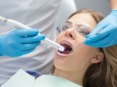
An intraoral camera is a compact, handheld imaging tool that captures crisp, full-color views from inside the mouth and displays them on a monitor in real time. Unlike a standard photograph, these cameras are designed specifically for oral anatomy—allowing dentists and hygienists to inspect teeth, restorations, gum tissue, and other soft tissues from angles that are otherwise difficult to visualize. The result is a detailed, magnified look that helps uncover issues that might be missed during a basic visual exam.
The device is small and gentle, typically shaped much like a pen, and the images it produces are high-resolution enough to show fine cracks, early enamel breakdown, staining, and the margins of crowns or fillings. Because images are immediate and color-accurate, they give both the clinician and the patient a shared reference point for discussion. This direct visual feedback can be especially helpful when explaining why a recommended treatment is appropriate.
Intraoral cameras are non-invasive and quick to use, so obtaining a full set of diagnostic images adds very little time to a routine visit. For patients who find dental procedures anxiety-provoking, the camera’s brief, painless imaging process is an efficient way to gather meaningful diagnostic information without introducing additional discomfort.
High-quality intraoral images extend the clinician’s ability to spot problems at an earlier stage. Subtle surface changes, tiny fractures, and beginning decay can be difficult to detect by sight or touch alone; magnified photography helps bring these details into focus. When paired with other diagnostic methods such as visual exams and digital radiography, intraoral cameras improve diagnostic confidence and can reduce the likelihood of overlooked concerns.
Because images can be viewed in real time, the clinician can adjust lighting, angle, and focus to examine suspicious areas from multiple perspectives, which supports more accurate clinical decisions. This is particularly valuable for assessing the integrity of existing restorations, monitoring wear patterns, and documenting suspicious tissue changes that warrant closer observation or biopsy.
Routine documentation with intraoral cameras also creates a baseline for comparison. When images are taken during regular visits, emerging patterns can be tracked over time—making it easier to identify gradual shifts in oral health and to intervene before conditions progress to more complex problems.
One of the most immediate benefits of intraoral cameras is the way they transform patient communication. Instead of relying solely on verbal descriptions, dentists can show patients exactly what they are seeing. This shared visual context helps patients better understand diagnoses, the condition of their teeth and gums, and the rationale behind treatment recommendations. Clear visual evidence often leads to more informed and comfortable decision-making.
For many patients, seeing an enlarged image of a problematic area removes ambiguity and builds trust in the care plan. The camera’s images make it easier to explain why certain steps—such as repairing a chipped filling, replacing a failing crown, or treating localized gum inflammation—are being suggested. Patients who are well-informed about their oral health can participate more actively in planning their treatment and maintenance routines.
Clinicians also use intraoral images as an educational tool during preventive visits. Imagery highlighting plaque buildup, areas missed during home care, or the early signs of enamel erosion provides concrete, actionable guidance that patients can apply at home to improve their daily oral hygiene.
Intraoral images can be saved directly into a patient’s electronic record, creating a permanent, date-stamped visual history of oral health. These records support continuity of care by allowing clinicians to review past images, compare progression, and make treatment adjustments informed by documented trends. Reliable documentation also reduces ambiguity during follow-up visits and helps maintain clinical accuracy across care episodes.
When collaborative treatment is required—such as a referral to a specialist or communication with a dental laboratory—high-quality intraoral images are invaluable. They allow specialists and technicians to assess the intraoral situation remotely, helping to streamline consultations and improve the precision of fabricated restorations. Clear imagery minimizes the need for repeated chairside adjustments and supports better outcomes in complex restorative or surgical cases.
Because images are digital, they can be shared securely with other providers when appropriate and with patient consent. This ease of sharing enhances coordinated care while preserving the detailed visual context that can be lost when relying on written notes alone.
Undergoing an intraoral camera exam is straightforward and comfortable. During a routine checkup or a focused evaluation, the dental professional will position the camera inside the mouth and capture several images of targeted areas. Each shot takes a moment, and the entire process rarely adds significant time to the appointment. Patients usually watch the images appear on-screen in real time, which creates an opportunity to ask questions as the clinician points out observations.
The process is non-invasive and free of radiation, since it relies on optical imaging rather than X-rays. Surfaces are examined at magnifications that reveal surface-level issues and soft-tissue changes, while X-rays continue to play a key role in assessing structures below the gum line. The camera and any accessories that come in contact with the oral tissues are cleaned and disinfected according to strict infection-control protocols to ensure patient safety.
Following an exam, images may be saved into the patient’s record for future comparison or included as part of a treatment plan illustration. Patients leave with clearer knowledge of their oral condition and a visual reference that reinforces the clinician’s recommendations. For those monitoring healing, tracking restorations, or following a preventive program, intraoral photos provide a tangible, visual timeline of progress.
At the office of Brian Howe DDS, Family Dentistry, intraoral cameras are one of several modern tools we use to deliver careful, patient-centered care. If you’d like to learn more about how intraoral imaging could help improve your diagnosis, treatment planning, or routine checkups, please contact us for more information.
An intraoral camera is a small, handheld imaging device designed to photograph the inside of the mouth with high-resolution, full-color detail. The camera captures magnified views of teeth, restorations and soft tissues and transmits those images to a chairside monitor in real time. Clinicians can adjust angle, lighting and focus to visualize surfaces and margins that are difficult to see with the naked eye.
The resulting images provide a precise visual record that supports diagnosis and treatment planning. Because the process relies on optical imaging rather than radiation, it is safe for routine use alongside other diagnostic tools. Images are often saved to the patient’s electronic chart for documentation and comparison over time.
Magnified, color-accurate images from an intraoral camera make subtle changes easier to spot, such as hairline cracks, early enamel breakdown and the margins of restorations. These details can be missed during a standard visual exam, so the camera supplements tactile and visual assessment to increase diagnostic confidence. When combined with digital radiography and a clinical exam, intraoral imaging helps clinicians detect problems earlier and with greater precision.
Early detection allows less invasive interventions and more predictable outcomes by addressing issues before they progress. Regular imaging creates a visual baseline that makes gradual changes easier to identify during follow-up visits. This documented progression supports informed clinical decisions and timely treatment when needed.
An intraoral camera exam is quick, noninvasive and generally well tolerated by patients of all ages. The device is typically pen-sized and designed for gentle intraoral use, so most images are captured without discomfort or extra chair time. Because the method uses optical imaging, it does not involve ionizing radiation and can be repeated as needed for documentation and monitoring.
Infection-control protocols require that any part of the camera that contacts oral tissues be cleaned or sheathed between patients. Clinicians also explain the process in real time while showing images on-screen, which helps reduce anxiety and invites patient questions. Overall, the procedure enhances comfort by providing visual clarity without adding invasive steps.
Seeing an enlarged image of a specific tooth or tissue gives patients a concrete visual reference that clarifies verbal explanations. When clinicians display images on a monitor and point out areas of concern, patients can more easily grasp the nature and urgency of recommended care. This shared visual context supports informed consent and encourages collaborative decision-making about treatment priorities.
Images can be annotated or grouped to illustrate progression, potential outcomes and the expected benefits of various interventions. Visual evidence often reduces ambiguity and increases patient comfort with proposed procedures. Educated patients are better equipped to follow home-care instructions and participate actively in their preventive plan.
Yes, intraoral images are typically saved directly into the patient’s electronic record as date-stamped documentation of oral health. These files create a visual history that clinicians can reference for treatment planning, follow-up care and continuity among providers. High-quality images also help specialists and dental laboratories understand the intraoral situation before a referral or restorative procedure.
When coordination with outside providers is necessary, images can be shared securely with patient consent to support remote consultations and laboratory communication. Clear visual context reduces ambiguity and helps minimize unnecessary repeat exams or chairside adjustments. Secure transfer and appropriate permissions are standard practice to protect patient privacy during collaboration.
Intraoral cameras capture surface and soft-tissue details in full color, while digital X-rays and CBCT reveal internal structures such as tooth roots and bone. Because each modality provides different information, combining them leads to a more complete diagnostic picture. Clinicians use camera images for surface assessment and patient education, and radiographs or CBCT for evaluating underlying anatomy and pathology.
This multimodal approach improves treatment planning by correlating visible surface changes with radiographic findings. For example, a discolored or cracked restoration can be evaluated visually with the camera and assessed for underlying issues with radiography. Coordinated interpretation of both types of images supports more accurate, predictable care.
During an exam, your dental professional will position the camera inside the mouth and capture several targeted images of teeth, restorations and soft tissues. Each shot takes only a moment, and patients typically see the images appear on the monitor in real time. The clinician may adjust angles and lighting to capture the clearest possible views of areas of interest.
The procedure rarely adds significant time to a routine visit and does not require special preparation. After imaging, selected photos may be saved to your record or used to illustrate a recommended treatment plan. Patients are encouraged to ask questions while viewing images to better understand their oral health status.
Routine imaging at successive visits creates a visual timeline that makes gradual changes easier to detect and quantify. Clinicians can compare date-stamped photos to observe trends such as restoration wear, enamel changes or soft-tissue healing. This documented progression helps determine whether conservative monitoring, intervention or revision is appropriate.
Consistent photographic records also support preventive conversations by showing areas of plaque accumulation or brushing technique issues. For patients undergoing restorative or cosmetic treatment, sequential images help verify healing and the longevity of work performed. Longitudinal documentation strengthens clinical decision-making and patient engagement in care.
Strict infection-control measures require that any camera components contacting oral tissues be either disinfected between patients or used with disposable barriers. Clinicians follow evidence-based cleaning protocols consistent with professional guidelines to maintain a safe environment. These steps minimize cross-contamination risk while allowing for routine intraoral photography.
On the privacy front, images stored in electronic records are protected by secure systems and access controls to comply with health information privacy standards. Sharing images with other providers is done only with patient consent and through secure channels. Together, these practices protect both patient safety and the confidentiality of visual records.
Intraoral cameras are useful at multiple stages of restorative and cosmetic care, including diagnosis, treatment planning and final verification. Clinicians use camera images to evaluate the condition of existing crowns, fillings and veneers, to communicate the desired aesthetic goals, and to document the appearance of preparations before and after treatment. The imagery helps laboratories fabricate restorations with a clearer understanding of margins, shade relationships and intraoral context.
During follow-up, intraoral photos can confirm fit, margin integrity and tissue response, reducing the need for repeated chairside adjustments. The visual record also aids long-term monitoring of restorative performance and esthetic stability. This consistent documentation supports precise, patient-centered outcomes in both restorative and cosmetic workflows.
Our friendly and knowledgeable team is always ready to assist you. You can reach us by phone at (740) 393-2161 or by using the convenient contact form below. If you submit the form, a member of our staff will respond within 24–48 hours.
Please do not use this form for emergencies or for appointment-related matters.
